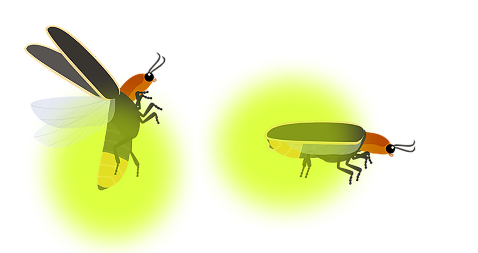
A Curtin University-led research team has found a way to synthetically create a firefly’s ‘glow’ that could have positive impacts on the access to medical light-imaging tools used to detect tumours and other diseases.
Fireflies emit their ‘glow’ due to a natural chemical reaction that happens in their abdomens in the presence of an enzyme called luciferase. The research team, through electrochemistry was able to trigger this same reaction without the enzyme, thereby artificially creating the same ‘glow’.
Lead researcher, Associate Professor Simone Ciampi, from the School of Molecular and Life Sciences said the breakthrough could increase use of this bioluminescent reaction, and reduce the costs of light bioluminescence imaging, which can be used to monitor diseases that lead to altered oxygen metabolism, such as cancer.
“Bioluminescence imaging that mimics the firefly’s glow is widely used in scientific research for mapping diseases, such as cancer, in animal tissues. A glow will indicate where oxygen metabolism is altered by the diseased cells,” Associate Professor Ciampi said.
“Currently thousands of fireflies are needed to extract small amounts of the enzyme – called luciferase –making firefly’s bioluminescence imaging very expensive and limiting its diagnostic uses to animals.
“Through our research, we have now found a way to recreate that firefly light electrochemically, triggering a reaction that uses superoxide, thereby making it cheaper and easier to harness and paving the way for use in human diagnostic tools.”
PhD student Mattia Belotti, from Curtin’s School of Molecular and Life Sciences, who also worked on the research, said the team also uncovered a way to change the colour of the light depending on the conditions and environment where the light-emitting reaction occurred.
“Through electrolyte engineering we were able to control the colour of the light to change from red to green where there were higher levels of oxidation, which typically occurs in a cell under stress from disease. In this situation, more green light means more ‘bad’ activity”, Mr Belotti said.
“This synthetic enzyme could also be used for detecting the formation of adenosine triphosphate (ATP), an organic compound responsible for providing energy to all living cells.
“Altered ATP metabolism (too much or too little energy available) is another marker that can be used to detect diseases in isolated cells and animal models. Identifying these defects early could be lifesaving as medical therapy and treatment approaches can be implemented quickly to help with disease regression.”
The research was an international collaboration between researchers at Curtin University, Flinders University, Australian National University and the University of Bologna.
The full paper titled ‘Luciferase-free Luciferin Electrochemiluminescence’ has been published in the Angewandte Chemie International Edition and is available here.